
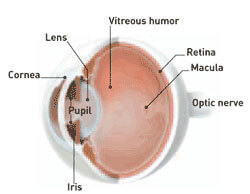
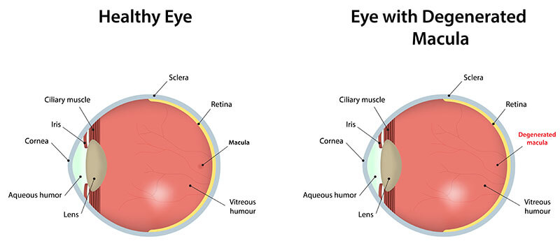
Macular degeneration is a deterioration or breakdown of the macula. The macula is a small area in the retina at the back of the eye that allows you to see fine details clearly and perform activities such as reading and driving. When the macula does not function correctly, your central vision can be affected by blurriness, dark areas or distortion. Macular degeneration affects your ability to see near and far, and can make some activities—like threading a needle or reading—difficult or impossible.
Although macular degeneration reduces vision in the central part of the retina, it usually does not affect the eye’s side, or peripheral, vision. For example, you could see the outline of a clock but not be able to tell what time it is.
Macular degeneration alone does not result in total blindness. Even in more advanced cases, people continue to have some useful vision and are often able to take care of themselves.
In many cases, macular degeneration’s impact on your vision can be minimal.
Many older people develop macular degeneration as part of the body’s natural aging process. There are different kinds of macular problems, but the most common is age-related macular degeneration (AMD). Exactly why it develops is not known, and no treatment has been uniformly effective. Macular degeneration is the leading cause of severe vision loss in Caucasians over 65.
Most people have the “dry” form of AMD. It is caused by aging and thinning of the tissues of the macula. Vision loss is usually gradual.
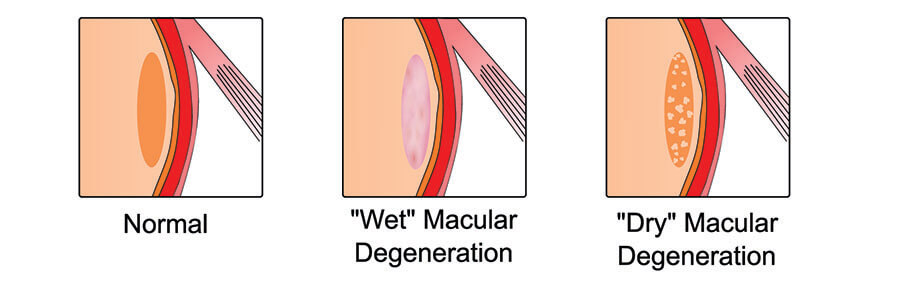
The “wet” form of macular degeneration accounts for about 10% of all AMD cases. It results when abnormal blood vessels form underneath the retina at the back of the eye. These new blood vessels leak fluid or blood and blur central vision. Vision loss may be rapid and severe.
Deposits under the retina called drusen are a common feature of macular degeneration. Drusen alone usually do not cause vision loss, but when they increase in size or number, this generally indicates an increased risk of developing advanced AMD. People at risk for developing advanced AMD have significant drusen, prominent dry AMD, or abnormal blood vessels under the macula in one eye (“wet” form).
Macular degeneration can cause different symptoms in different people. The condition may be hardly noticeable in its early stages. Sometimes only one eye loses vision while the other eye continues to see well for many years.
But when both eyes are affected, the loss of central vision may be noticed more quickly.
Following are some common ways vision loss is detected:
Many people do not realize that they have a macular problem until blurred vision becomes obvious. Your ophthalmologist (Eye M.D.) can detect early stages of AMD during a medical eye examination that includes the following:
Although the exact causes of macular degeneration are not fully understood, antioxidant vitamins and zinc may reduce the impact of AMD in some people.
A large scientific study found that people at risk for developing advanced stages of AMD lowered their risk by about 25% when treated with a high-dose combination of vitamin C, vitamin E, beta carotene and zinc. Among those who have either no AMD or very early AMD, the supplements did not appear to provide an apparent benefit.
It is very important to remember that vitamin supplements are not a cure for AMD, nor will they restore vision that you may have already lost from the disease. However, specific amounts of these supplements do play a key role in helping some people at high risk for advanced AMD to maintain their vision. You should speak with your ophthalmologist to determine if you are at risk for developing advanced AMD, and to learn if supplements are recommended for you.
“Wet macular degeneration can be treated with injections of medication into the eye, photodynamic therapy (PDT), or laser surgery, injections of anti-VEGF agents such as Lucentis or Avastin are currently first-line treatments for “wet” macular degeneration. These medications reduce the growth of abnormal blood vessels and slow their leakage.
If the “wet” macular degeneration is not responding well to the injections of medications into the eye, the photodynamic (PDT) can be used in combination with the injections. PDT uses a combination of special drug and laser treatment to slow or stop leaking blood vessels. Lasers surgery, a brief outpatient procedure that uses a focused beam of light to slow or stop leaking blood vessels that damage the macula, also can be used in combination with the injetions of medication into the eye.
All of these treatments are performed at your doctor’s office.
These procedures may preserve more sight overall, though they are not cures that restore vision to normal. Despite advanced medical treatment, many people with macular degeneration still experience some vision loss.
To help you adapt to lower vision levels, your ophthalmologist can prescribe optical devices or refer you to a low-vision specialist or center. A wide range of support services and rehabilitation programs are also available to help people with macular degeneration maintain a satisfying lifestyle. Because side vision is usually not affected, a person’s remaining sight is very useful. Often, people can continue with many of their favorite activities by using low-vision optical devices such as magnifying devices, closed-circuit television, large-print reading materials and talking or computerized devices.
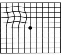
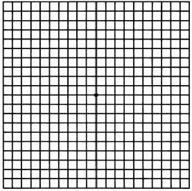
You can check your vision daily by using an Amsler grid like the one pictured here. You may find changes in your vision that you wouldn’t notice otherwise. Putting the grid on the front of your refrigerator is a good way to remember to look at it each day.
For more information, please contact:
The American Academy of Ophthalmology
(415) 561-8500
www.aao.org
The American Macular Degeneration Foundation
(413) 268-7660
www.macular.org
Foundation Fighting Blindness
(888) 394-3937
www.blindness.org
You’ll see that when it’s time to make an appointment with a specialist in Retina and Vitreous diseases, you can trust your eye health to the team at Inland Valley Retina Medical Associates, Inc.
If you would like to speak with the Corporate Administrator, please contact Lee Barsdorf
Phone: 951.679.0400
Fax: 951.672.6667